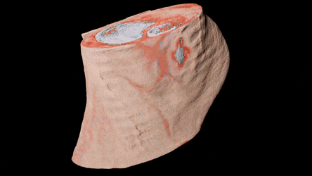
Stunning new color X-ray pictures, from a corporation referred to as Mars Bioimaging, in New Seeland, appear to create flesh and bone semitransparent and hyperreal.
The gif on top of shows one among the company's strange and interesting images: a slice of human articulatio talocruralis, with off-white, rugged bones, bloody-looking muscle tissue and a pad of fat dirty giving protection below the heel with a whipped-cream texture.
This image shows a articulatio plana with a lot of muscle, less visible bone, nearly no fat and a clearly-articulated watch:
It's important to notice that these are not "true-color" X-ray scans as the general public would unremarkably perceive the term. because the inventors of the sensing element that was accustomed create these pictures delineate in an exceedingly 2015 paper within the journal IEEE Transactions on Medical Imaging and on the company's web site, the colours in these pictures ar applied supported the computer's detection totally different|of various} wavelengths of X-rays passing through different substances. There are, however, no "true" red X-rays or "true" white X-rays; the device's programmers assign completely different|completely different} colours to different detected body components. (What human brains interpret as color comes from completely different wavelengths of sunshine within the visual spectrum bouncing off objects. lightweight|light|visible radiation|actinic radiation|actinic ray} is additionally a variety of radiation however is lower-energy than X-ray light.)
To with success distinguish muscle, fat and bone, Mars Bioimaging developed sensors that might work within CAT (CT) scanners (circular X-ray devices that manufacture three-dimensional X-ray images) and manufacture terribly careful info concerning the wavelengths of individual X-ray photons that withstand and bounce off human tissue. By sensing the wavelengths that disappear once passing through a selected little bit of tissue, the device makes a judgement concerning what chemicals structure that tissue and uses that info to work out what kind of tissue it had been. The photon-counting technology, the corporate says in its promoting materials, was originally developed as a part of its founders' work with CERN, the eu Organization for Nuclear analysis, that operates the world's largest scientific instrument.
By matching those scans with details regarding however completely different chemical compounds act with X-ray lightweight, they were able to distinguish completely different compounds in X-ray scans, the researchers wrote within the 2015 study. to provide these new grody, beautiful color pictures of living tissue, they merely tasked the pc with painting the various compounds of fat, bone and muscle completely different colours.
The profit for researchers, the corporate claims in its selling materials, is not most the fascinating visuals (though that is a plus) because it is that the wealth of precise chemical knowledge on objects within the scanner. The careful, multilayered tissue scans, they write, can change new exactitude in medical analysis.
Originally published on Live Science.


Congratulations! This post has been upvoted from the communal account, @minnowsupport, by freebook from the Minnow Support Project. It's a witness project run by aggroed, ausbitbank, teamsteem, theprophet0, someguy123, neoxian, followbtcnews, and netuoso. The goal is to help Steemit grow by supporting Minnows. Please find us at the Peace, Abundance, and Liberty Network (PALnet) Discord Channel. It's a completely public and open space to all members of the Steemit community who voluntarily choose to be there.
If you would like to delegate to the Minnow Support Project you can do so by clicking on the following links: 50SP, 100SP, 250SP, 500SP, 1000SP, 5000SP.
Be sure to leave at least 50SP undelegated on your account.
Thank you for comment my post