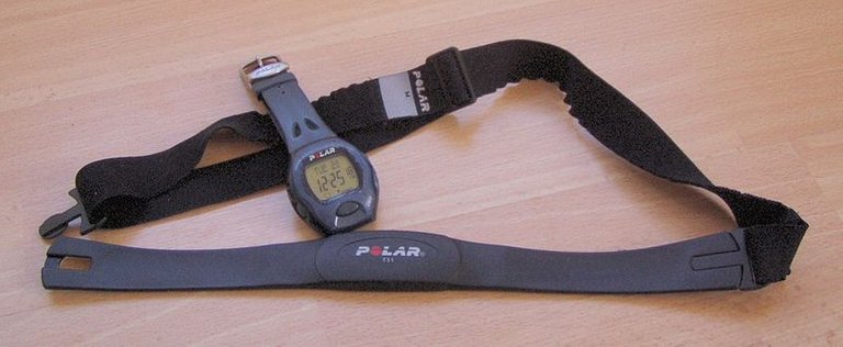
Heart rate is modified according to the needs of the body. The heart increases during physical exercise to supply extra oxygen to the tissues and to remove excess carbon dioxide. At rest, a normal adult human heart beats about 75 times per minute; but during very tedious exercise it might beat 200 times per minute. Heart rate is controlled by the Sinoatrial Node (SAN). The rate increases or reduces when it receives information via two autonomic nerves that link the SAN with the cardiovascular centre in the medulla of the brain.
- A sympathetic or accelerator nerve speeds up the heart. The synapses at the end of this nerve secrete noradrenaline.
- A parasympathetic or decelerator nerve, a branch of the vagus nerve, slows down the heart. The synapses at the end of this nerve secrete acetylcholine.
A negative feedback system functions to control the level of carbon dioxide and, conversely, oxygen in the blood. During exercise, the level of carbon dioxide starts to increase. This is detected by chemoreceptors (cells sensitive to chemical change) situated in three places: the carotid artery, the aorta and the medulla. Impulses travel down the sympathetic nerve and heart rate increases. When the carbon dioxide level drops low enough, impulses pass down the parasympathetic nerve, and the heart rate returns to normal. The control of heart rate is closely related to the control of breathing.

Several factors affect heart rate:
- Secretion of adrenaline (epinephrine) in response to stress, excitement or other emotions. Adrenaline is the hormone that prepares the body for action, and one of its effects is to increase heart rate.
- Movement of the limbs, as in exercise. It is thought that stretch receptors in the muscles and tendons relay information to the brain, telling the cardiovascular centre that oxygen levels will soon fall and that carbon dioxide will soon build up. This initiates signals that increase heart rate and breathing rate.
- The level of respiratory gases in the blood, as described above.
- Blood pressure. When blood pressure gets too high, a fail-safe mechanism prevents any further increase in heart rate.
BLOOD VESSELS
There are three types of blood vessels: arteries, veins and capillaries. The structure of each is closely related to its function.
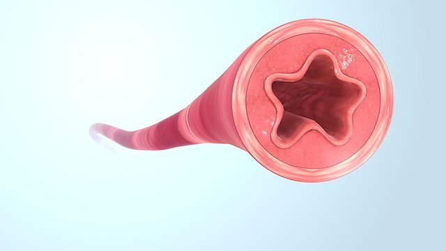
Arteries carry blood away from the heart towards other organs of the body. Arteries branch into smaller arterioles, which branch into tiny capillaries. These are permeable (‘leaky’) vessels whose walls are one cell thick to allow the exchange of materials between blood and nearby cells. Blood flows from capillaries into venules, which drain into larger veins. The walls of arteries and vein have the same three layers: the tunica externa, tunica media and tunica intima or interna. The relative thickness and composition of this layers vary according to the function of the vessel. The artery, which has to withstand pulses of high pressure, has an especially thick tunica media containing smooth muscle and elastic fibres. Veins have a thinner tunica media and a larger lumen to carry slower-flowing blood at low pressure. Valves in veins prevent blood flowing backwards.

CAPILLARY CIRCULATION
The circulatory system keeps all cells bathed in tissue fluid. All living cells are surrounded by tissue fluid. This is also known as interstitial or intercellular fluid. The tissue fluid has a reasonably constant composition because permeable capillaries allow exchange of materials between the fluid and the blood.
How tissue fluid is formed
Blood from the arterioles is under high hydrostatic pressure. When blood enters a capillary, substances are forced out through the permeable capillary wall. Hydrostatic pressure is physical pressure. The contraction of the ventricles of the heart creates hydrostatic pressure in the vessels. The capillary walls act as filters and a proportion of all chemicals below a particular size is squeezed out, forming tissue fluid. The composition of tissue fluid is very similar to that of plasma, but tissue fluid lacks most of the large proteins that cannot pass through the capillary wall. Plasma proteins remain in the blood, where they play an important part in the drainage of tissue fluid.
STRUCTURE AND FUNCTION IN BLOOD VESSELS
It is important to relate the structure of different blood vessels with their function. Arteries have tough elastic walls that withstand the high pressure of blood that flows through them as it is forced out of the heart. Blood pressure is at its highest in the large arteries supplied by the left ventricle. The pressure then reduces as blood passes through the smaller arterioles and then into the capillaries. The rate of blood flow is slowest through the capillaries, which also have the largest surface area of the entire blood vessel system. This is because this is where gas exchange occurs. Blood flow then increases as blood enters the veins to travel back to the heart. Veins carry blood at low pressure, so their walls are thin and have a large lumen. The valves prevent backflow.
How tissue fluid returns to the blood
Two main forces act on the blood as is it passes along the capillary; the first is the hydrostatic pressure of the blood: this tends to force water and solutes out of the capillary. The second is the water potential of the plasma; which tends to draw water into the capillary by osmosis.
As blood flows along a capillary, fluid passes to the tissues, and the volume and hydrostatic pressure of the blood in the capillary decrease. Cell waste, such as urea, and substances that have been secreted into the tissue fluid, diffuse into the capillary, contributing to the composition of venous blood.
BLOOD FLOW IN VEINS
Blood that drains into the venules is deoxygenated, under low pressure and contains many waste products and cell secretions. Several features allow blood to return to the heart:
- Valves in veins close and prevent backflow.
- Working muscles surrounding the veins squeeze blood along as they contract. The action of muscles, especially those in the legs, is so important that it has been called the ‘secondary heart’ or the ‘venous pump. This venous pump is very important to the circulation, and regularly standing still for any length of time can lead to problems. Millions of people suffer from varicose veins or from haemorrhoids (piles), damaged veins whose walls have been stretched by pools of accumulated blood.
- Gravity helps blood to flow from organs ‘above’ the heart.
- The negative pressure created in the thorax during inspiration (breathing in) draws blood from surrounding veins.
- The action of the heart. After systole (contraction), the elastic walls of the chambers recoil, drawing blood in from the veins.
CONTROL OF BLOOD FLOW
The body often needs to alter blood flow to different areas, according to circumstances. For example, when we are hot, we lose excess heat because more blood flows to the surface of the skin. Blood flow can be modified by sphincters and shunt vessels. A sphincter is a ring of muscle around a blood vessel which can contract, reducing the lumen size of the vessel and so reducing or preventing blood flow to a particular area. Sphincters can redirect blood into shunt vessels and these bypass a particular area. For instance, when we are cold, sphincters reduce blood flow to the skin surface, redirecting it along shunt vessels that keep the blood deeper in the body.
Blood flow can also be modified by altering the diameter of the vessel itself. Many blood vessels, particularly arterioles, contain smooth muscle fibres that can contract to reduce blood flow through the vessel; this is called vasoconstriction. Blood flow increases when the muscle fibres relax; this is vasodilation. Blood pressure can be regulated by altering the degree of vasoconstriction or vasodilation.
THE LYMPHATIC SYSTEM
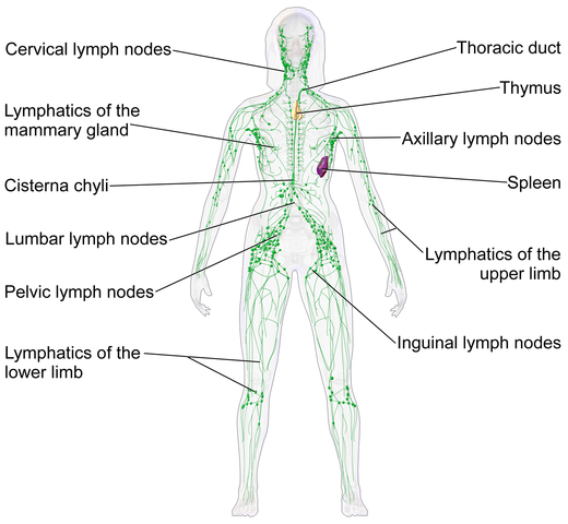
The lymphatic system is part of the immune system and also part of the circulatory system: it returns to the heart the small amount of tissue fluid that cannot be returned by the veins.We can think of the lymph system as an extra set of veins. Flow through lymphatic vessels is slow, but important. Lymphatic flow begins in the areas round blood capillaries (capillary beds), where small amounts of tissue fluid drain into tiny lymphatic capillaries. The walls of these vessels are more permeable than blood capillaries to lipids and large molecules such as proteins.
This is why lymph contains a high proportion of these substances. Many cells secrete substances that are too large to enter the blood directly, and these substances pass into the general circulation via the lymphatics. The lymph capillaries drain into larger lymph vessels that look like thin, transparent veins. These vessels have valves to prevent backflow. Lymph contains no red blood cell, and so is pale and clear. All lymph vessels flow towards the upper thorax, passing through numerous lymph nodes that filter out bacteria and cell debris.

BLOOD PRESSURE
The term blood pressure refers to the physical pressure of blood flowing through the arteries. This depends on the force created by the pumping of the heart, the volume of circulating blood and the size of the blood vessels. The body must keep blood pressure inside fairly strict limits. It must be high enough to force blood through all the capillaries so that all cells are well nourished, but not high enough to put the heart and blood vessels under strain: high blood pressure causes various health problems.
In the measurement of blood pressure, the diastolic pressure is used as an indicator for medical problems: a normal reading is between 60 and 80 mmHg, with anything over 90 usually regarded as high.
Blood pressure is usually measured using a sphygmomanometer. An inflatable cuff is placed around the upper arm and inflated until all blood flow, in or out of the arm, stops. The blood flow in the brachial artery (at the elbow) is monitored using a stethoscope. After inflation, there is no sound, but, as air escapes from the cuff, the pressure decreases until it falls just below that created by the heart as it contracts. At this point, blood is heard spurting through the constriction in the artery. This pressure is the systolic value. Pressure in the cuff continues to drop until blood can be heard flowing constantly. This is the diastolic value and represents the pressure to which arterial blood falls between beats
Have you ever stood up very quickly, and then felt faint or dizzy? This happens because the rapid change in posture has altered the body’s blood distribution, causing a temporary lack of blood to the brain. Fortunately, body mechanisms quickly bring the blood flow back to normal.
CONTROL OF BLOOD PRESSURE
A negative feedback system keeps blood pressure inside safe limits. The pressure of the blood is detected by the carotid sinus, a small swelling in the carotid artery. When blood pressure gets too high, the walls of the sinus expand and stretch receptors in the carotid artery wall send information to the cardiovascular centre in the medulla. Signals from the medulla then lower heart rate and cause vasodilation, so lowering blood pressure.
Many mechanisms work together to keep blood pressure constant. The size of blood vessels, the heart rate and the volume of circulating blood can all change to increase or decrease blood pressure, according to the needs of the body. The kidneys are particularly important because they control how much fluid we lose. A drop in blood volume and pressure causes more fluid to be retained in the blood and less to be lost in the urine.
Thank you for reading.
REFERENCES
- All about heart rate pulse
- heart rate
- Blood vessel
- Blood vessel
- blood physiology
- Capillary Circulation
- Blood vessel structure and function
- Blood flow and blood pressure regulation
- Local blood flow regulation
- ncbi
- Lymphatic system
- Lymphatic system
- High blood pressure hypertension
- Understanding blood pressure
- Control-of-blood-pressure
Thanks for your contribution to the STEMsocial community. Feel free to join us on discord to get to know the rest of us!
Please consider supporting our funding proposal, approving our witness (@stem.witness) or delegating to the @stemsocial account (for some ROI).
Thanks for using the STEMsocial app
, which gives you stronger support. Including @stemsocial as a beneficiary could yield even more support next time.