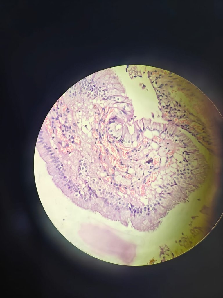
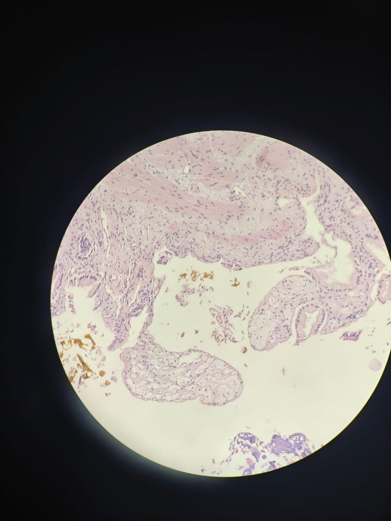
Hello everyone, how are you all? Hope you all are doing well and i am too. Today we are going into gall bladder specimen. A gall bladder specimen came and grossing was done and few yellow streak lines are noted grossly and then from from head, neck and fundus sections were taken. After all processing slides were given. When slides are seeing under microscope then i found these features.
What is cholesterolosis?
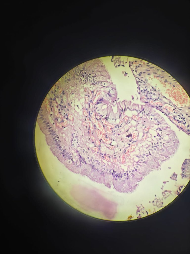
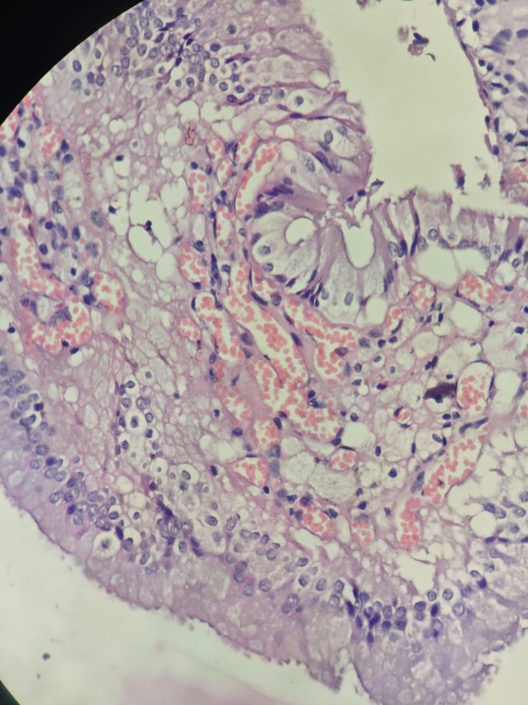
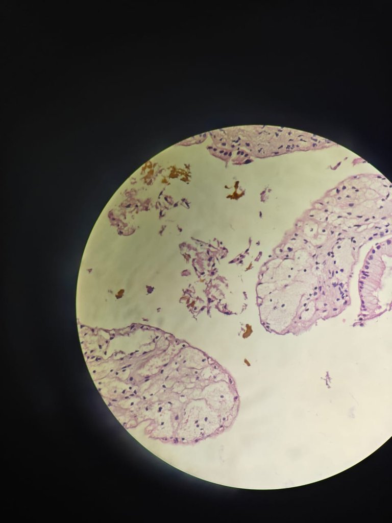
In the subpithelium(below the epithelial lining) it is called lamina propria and there is accumulation of lipids which includes triglycerides, cholesterol precursors and cholesterol esters in the macrophages of that lamina propria.
These macrophages ppear foamy because of this accumulation of lipids inside them. It is also called as strawberry gall bladder.
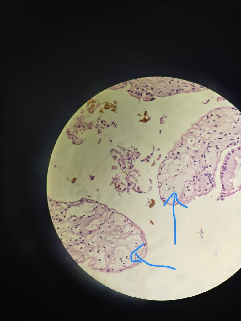
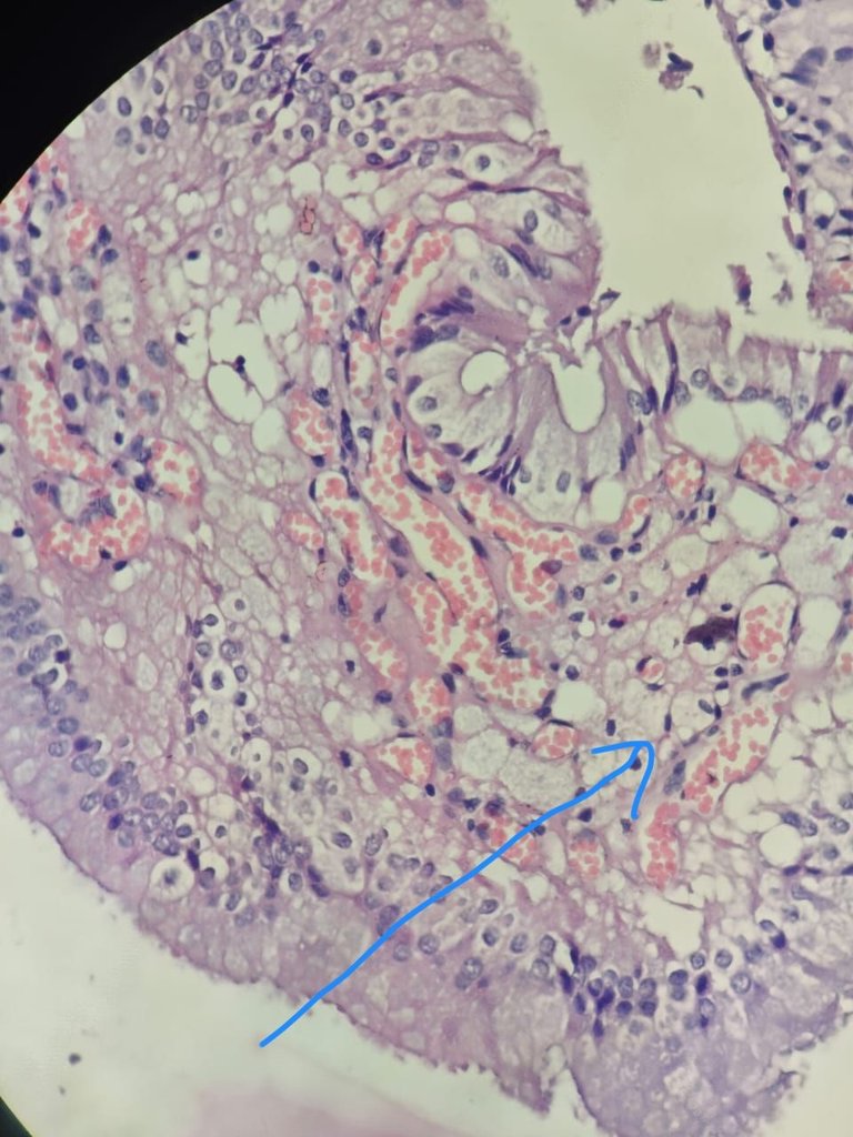
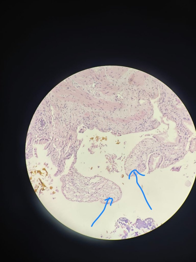
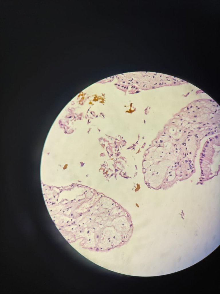
In the above images, you can see i labelled with blue arrows which are showing foamy macrophages in the lamina propria.
This is not neoplastic condition. Its most commonly seen condition and infact 2nd most commonly seen after cholelithiasis. So no need to worry about this thing. This is due to if we have high Body Mass Index(BMI) and in females. Just be aware about your diet what you are taking daily and do exercise everyday.
I just wants to show this entity to you once so that you see this one and when you see pathology report also you will come to know what it is.
References
- Rosai and Ackerman's Surgical Pathology - 11th Edition
Thanks for reading,
With regards,
Thanks for your contribution to the STEMsocial community. Feel free to join us on discord to get to know the rest of us!
Please consider delegating to the @stemsocial account (85% of the curation rewards are returned).
You may also include @stemsocial as a beneficiary of the rewards of this post to get a stronger support.
Congratulations @salomijale! You have completed the following achievement on the Hive blockchain And have been rewarded with New badge(s)
Your next target is to reach 700 upvotes.
You can view your badges on your board and compare yourself to others in the Ranking
If you no longer want to receive notifications, reply to this comment with the word
STOPThe white bulging appearance indicates lipid compartment. Thanks for the education.