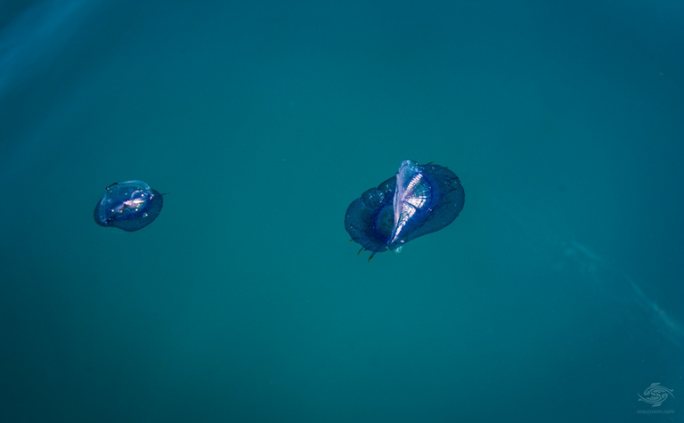
The “SAILING FLEET” of the intestinal cavities The class of hydroids is divided into two subclasses - hydroids and siphonophores. We pass on to the description of these amazing pelagic colonial intestinal cavities. The whole world of living beings lives on the verge of two elements - water and air. On floating algae, fragments of wood, pieces of pumice and other objects, one can find a variety of animals that have grown or firmly clung to. It should not be thought that they got here by chance - "in distress." On the contrary, many of them are closely connected with both the water and the air environment, and in other conditions they cannot exist. In addition to such "passive passengers", here you can also see animals actively swimming near the surface itself, equipped with variously arranged organs - floats, or animals that are held using a film of surface tension of water. This whole complex of organisms (pleuston) is especially rich in the subtropics and tropics, where the destructive effect of low temperatures is not felt. Above, when it came to the action of stinging cells, the “Portuguese warship” was already mentioned - a large physalia siphonophore (Physalia, see color table 8). Like all siphonophores, physalia is a colony, which includes both polypoid and medusoid individuals. An air bubble, or pneumatophore, rises above the surface of the water, a modified medusoid individual of the colony. In large specimens, the pneumatophore reaches 30 cm, it usually has a bright blue or reddish color. An air bubble floats on the surface of the sea like a tightly inflated rubber balloon. The gas that fills it is close in composition to air * but differs in an increased content of fuel oil and carbon dioxide and a reduced amount of oxygen. This gas is produced by special gas glands located inside the bubble. The walls of the pneumatophore withstand quite strong gas pressure, since they are formed by two layers of ectoderm, two layers of endoderm and two layers of mesoglea. In addition, the ectoderm secretes a thin chitinoid membrane, due to which the strength of the pneumatophore also increases significantly, although its walls remain very thin. The upper part of the pneumatophore has an outgrowth in the form of a comb. The comb is located on the pneumatophore somewhat diagonally and has a slightly curved S-shape. Other. 
seaunseen.com