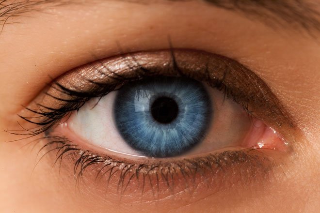
THE EYE

Although OOU exam is still in progress, I just want to drop this before I start reading for the next paper.
“Dad is speaking with his two sons (Charles and Simon); he is trying to know their future ambition.”
Dad: Charles, what will you like to become in future?
Charles: Dad, I would like to become a medical doctor
Dad: OK, that is a good idea; we all know the position of doctors in the world. What of you Simon?
Simon: Dad, I want to become an Optician
Charles: What!!! Optician?
Simon: Yes of course
Dad: Why do you want to become an optician, you don’t even think of any other work like Engineering, Pharmacy etc.
Simon: There is only one major reason why I am willing to be an optician and the reason is because of you and other people across the world
Dad: Me?
Simon: *Yeah dad, You know, I normally wonder what happen whenever you complain to mum about your eyes and to this reason I am gonna become an optician so I would find solutions to all the problem both on your eyes and others across the world.
(We will be speaking on sense organs and today’s topic will be based on sensation of vision, as we all know that whenever we speak about vision we are referring to the eyes and eye is an important part that someone must not play with).
SPECIAL SENSES
Special senses or special sensation are broad sensation that have specialized organs. The special senses are different from somatic senses that come from the skin, joint, muscles etc.
Medically and anatomically we have four senses which are;
- Sensation of vision
- Sensation of hearing and balance
- Sensation of smell
- Sensation of taste
well labelled
EYE
The eyeball is a globular organ with a diameter of 24mm. It is made up of two segments which are the upper part and the lower part. The upper part is small and it covers one-sixth of the eyeball while the lower part is large and it covers the rest five-sixth of the eyeball.
LAYERS OF THE EYEBALL
The eyeball is divided into three layers, which are the following;
A. The outer layer (Tunica Externa):
The Cornea is the outer part of the eyeball and it’s the clear dome part of the eye at the front part of the eye, it is a fibrous layer and comprise of two structures; the Iris and the Pupil. The cornea looks like a window in the eye.
Layers of the Cornea
- The corneal epithelium
- Bowman’s layer
- The corneal stroma
- Descemet’s
- The corneal endothelium
B. The middle layer (Tunica vasculosa):
The choroid makes up the middle layer of the eye, it is a thin vascular layer and it lies in between the sclera and the retina. It supplies blood to the utter retina and even provides 90% of the blood in the eye.
Layers of Choroid
- Haller’s layer
- Sattler layer
- Choriocapillaries
- Brunch membrane
C. Inner layer (Tunica interna):
The Retina is a light sensitive membrane that forms the inner part of the eye. Retina is a thin layer of the cells located at the back of the eyeball. The presence of nerve cell in it makes it sensitive to light.
Layers of thee Retina
- Pigment epithelium layer
- Rods and cones layer
- External limiting membrane
- Outer plexiform layer
- Inner plexiform layer
- Inner nuclear layer
- Outer nuclear layer
- Nerve fiber later
- Ganglion cell layer
- Internal limiting membrane
IMAGE FORMING MECHANISM
Whenever we look at an object, there is always a ray coming from the object and the ray is refracted by the eye lens and brought to a focus upon the retina. The image of the object then fall on the retina in an inverted direction and it’s been reversed from one side to another side. The cerebral cortex now helps in seeing the image in an upright position.
The eye muscles

MUSCLES OF THE EYE
The muscles of the eye are of two types;
i. Intrinsic muscles.
ii. Extrinsic muscles.
The intrinsic muscles: These are the muscles that are built up by the autonomic system and they are:
a. Constrictor papillae
b. Dilator papillae
c. Ciliary muscle
The extrinsic muscles: These are the muscles that are formed by the skeletal muscle and are controlled by the somatic nerves. The following are the muscles that surround the eye, though they are small and they are not that strong but they perform important work by allowing the eyes to carry out some specific task like scanning of object, maintaining stable image, tracking moving object etc.
- Inferior oblique muscle
- Superior oblique muscle
- Superior rectus muscle
- Inferior rectus muscle
- Lateral rectus muscle
- Medial rectus muscle
INNERVATIONS OF THE EYE
The eye is innervated by the somatic nerve fibers (the somatic nerve fiber include; Sympathetic and Para-sympathetic nerve fiber) and the autonomic nervous system. Generally the eye is innervated by;
- Sympathetic nerve fiber
- Para-sympathetic nerve fiber
- Oculomotor nerve
- Trochlear nerve
- Abducent nerve
FLUIDS OF THE EYE
The upper segment of the eye contains a fluid called the intraocular fluid. This fluid flow out via the pupil and also it is responsible for the maintenance of the eye shape. The intraocular fluid is divided into two viz.
a. Vitreous humor
b. Aqueous humor
Vitreous humor: This is a viscous fluid that is clear like a gel and it is composed of water and it is located in between the lens and the retina. Its major function is to give shape to the eye.
Aqueous humor: This is a transparent/thin fluid that is similar to plasma and it is located in the front of the retina. The aqueous humor is divided into anterior and posterior chamber and the two chambers communicate with each other via pupil.
Properties of the aqueous humor
Volume: 0.13ml
Reaction: Alkaline
pH: 7.5
Viscosity: 1.029
Refractory index: 1.34
Composition of the aqueous humor
- Water: 98.7%
- Solid: 1.3%
The solid is further divided into two;
- Organic
a. Albumin
b. Globulin
c. Glucose
d. Pyruvate
e. Lactase
f. Urea - Inorganic
a. Potassium
b. Phosphate
c. Chloride
d. Magnesium
e. Calcium
f. Sodium
g. Bicarbonate
THE EYE LENS
The eye lens is crystalline in nature and it’s located behind the pupil. It is a transparent biconcave and elastic structure in the eye; it helps in refracting light to the focused on the retina. The focal length of human eye lens is 44mm.
Structures of the eye lens
- Lens substances: Lens is formed by the lens fiber which are from the anterior epithelium and the fibers which are prismatic in nature.
- Anterior epithelium: This is a single layer of cuboidal epithelial cells; it’s located under the capsules.
- Capsule: This is the elastic membrane that covers the eye lens.
Applied physiology
GLAUCOMA
Glaucoma is a disease of the eye that is caused by increase in intraocular pressure; it causes damage of the optic nerve which results in blindness.Glaucoma is divided into two types; the primary open-angle glaucoma which is the most common type and the primary angle closure glaucoma.
CATARACT
This is the cloudiness of the eye lens, i.e. it makes the eyes to look doll even during the day and ray of light won’t be able to pass through the eye lens. It majorly causes blindness all over everywhere in the world. It is an old age disease that is developed after 55 years.
Conclusion:
Eyes is the light of the body; they play an important role in our day to day activities, anyone without eyes is totally in darkness, so therefore make sure your eyes is in good conditions.
Watch out for the other part of the sense organs, I believe it’s going to be more interesting. Kindly up-vote, resteem and follow me @timileyin988
Further readings
https://en.wikipedia.org/wiki/Extraocular_muscles
http://www.innerbody.com/anatomy/muscular/head-neck/muscles-eye
https://www.glaucoma.org/glaucoma/anatomy-of-the-eye.php

very nice post technical about eyes good for researches
are u a doctor?
I am a student physiologist
Nice post . good luck on physiologist studying
Eye, like your post!😄
Nice post, you listed alot of information about the eye, this reminds me of the last time i went to the eye doctor haha. They have these X-ray machines that save an image of your whole eye, freaky! Just glad i have perfect vision. Enjoyed Reading your post, tons of facts here! Have a Good Day/Evening.
Keep up The Excellent Content!
🐠Steem-on!🐟 @timileyin988