In one of my previous posts I discussed the potential of using mRNA instead of plasmid DNA for gene therapy applications. Here, I wanted to discuss in greater detail what are the hurdles of such approach and what scientists are doing to overcome them.
Lately, we learned so much about how a cell can decode the information contained in its DNA and use it to make proteins. Kids nowadays even study it in school so I’m pretty sure you have all heard of mRNA and you probably know that it’s the link between DNA and proteins. This molecule is so powerful that cells had to devise several ways to destroy it rapidly, otherwise, its prolonged presence in the cell could create havoc. The proteins capable of destroying RNA are called RNAses and because they are very stable, they can survive even outside the cells and they are basically everywhere in our environment. Therefore, when RNA is outside the cells it can degrade very fast.
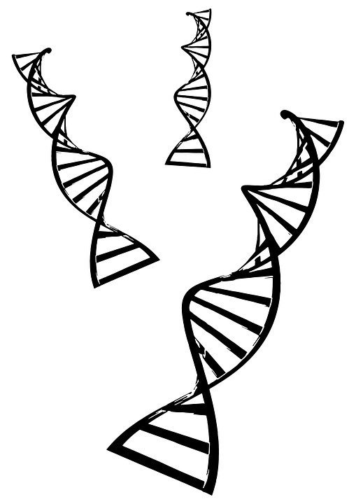
Image CCO Creative Commons Source
This is very problematic for handling and storing mRNA. So, we have identified the first issue with RNA, it’s not stable. Already in the 1970s, scientists were able to use mRNA to induce the expression of therapeutic proteins in different cell models (Gurdon et al. 1971; Laskey, Gurdon, and Crawford 1972). However, its poor stability prevented its adoption in clinical applications for several years. Another problem is that, to be expressed, mRNA needs to be delivered in the cytoplasm of a cell and because both mRNA and cell membrane are negatively charged, the mRNA on its own is not able to enter the cells. It was estimated that only 1 mRNA molecule every 10,000 eventually makes it inside a cells (Sahin, Karikó, and Türeci 2014).
Today, we are able to overcome mRNA stability and delivery issues by using lipidic or polymeric systems that have high affinity for the mRNA. They bind it forming particles called complexes. Because these complexes are positively charged they can enter the cells where they release their mRNA cargo. Complexes fulfil also a protective role because they can shield mRNA from RNAses, increasing its half-life. While all this sounds wonderful, there is another issue that prevents mRNA adoption in the clinic, mRNA is highly immunogenic. Because mRNA is used also by several pathogens like viruses to infect cells, our evolution provided us with several sensors that can detect mRNA. When mRNA is found in places where it should not be, these sensors can trigger an immune response. To be precise, some of these RNA sensors are: Toll-like receptors, RIG-1 and PKR (Alexopoulou et al. 2001; Diebold 2004). For this reason, for years the only application where mRNA was used was the development of vaccines, where immune-stimulation is required.
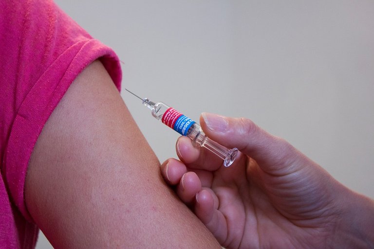
Image CCO Creative Commons - Source
However, in 2008 some scientists found that by exchanging all the uridines with pseudouridines they could reduce greatly the immunogenicity of mRNA (Karikó et al. 2008). This lead to the adoption of mRNA to direct the translation of therapeutic proteins in vivo. The increase in the number of scientific studies deploying mRNA led to further discoveries like the most recent that shows that there are alternative ways to decrease the immunogenicity of mRNA. One such approach consists in engineering the sequence of mRNA to incorporate the most GC-rich codons for each amino acid. By increasing the GC-content of the mRNA sequence, these scientists decreased the overall uridine content, drastically decreasing the immunogenicity of the mRNA without resorting to modified nucleosides (Thess et al. 2015). Because of these advances, today it is relatively easy to manufacture synthetic mRNA in vitro and deliver it effectively inside cells. This is a great tool at our disposal because it enables us to easily design custom mRNA sequences that can instruct cells to produce therapeutic proteins.
References
Alexopoulou, L, A Czopik Holt, R Medzhitov, and R A Flavell. 2001. “Recognition of Double-Stranded RNA and Activation of NF-Kappa B by Toll-like Receptor 3.” Nature 413(6857): 732–38.
Diebold, S. S. 2004. “Innate Antiviral Responses by Means of TLR7-Mediated Recognition of Single-Stranded RNA.” Science 303(5663): 1529–31. http://www.sciencemag.org/cgi/doi/10.1126/science.1093616.
Karikó, Katalin et al. 2008. “Incorporation of Pseudouridine into MRNA Yields Superior Nonimmunogenic Vector with Increased Translational Capacity and Biological Stability.” Molecular Therapy 16(11): 1833–40.
Gurdon, J B, C D Lane, H R Woodland, and G Marbaix. 1971. “Use of Frog Eggs and Oocytes for the Study of Messenger RNA and Its Translation in Living Cells.” Nature 233(5316): 177–82. http://www.ncbi.nlm.nih.gov/pubmed/4939175.
Laskey, R A, J B Gurdon, and L V Crawford. 1972. “Translation of Encephalomyocarditis Viral RNA in Oocytes of Xenopus Laevis.” Proceedings of the National Academy of Sciences of the United States of America 69(12): 3665–69. http://www.ncbi.nlm.nih.gov/pubmed/4345506.
Sahin, Ugur, Katalin Karikó, and Özlem Türeci. 2014. “MRNA-Based Therapeutics-Developing a New Class of Drugs.” Nature Reviews Drug Discovery 13(10): 759–80. http://dx.doi.org/10.1038/nrd4278.
Thess, Andreas et al. 2015. “Sequence-Engineered MRNA Without Chemical Nucleoside Modifications Enables an Effective Protein Therapy in Large Animals.” Molecular Therapy 23(9): 1456–64. http://dx.doi.org/10.1038/mt.2015.103.
Wolff, J A et al. 1990. “Direct Gene Transfer into Mouse Muscle in Vivo.” Science (New York, N.Y.) 247(4949 Pt 1): 1465–68. http://www.ncbi.nlm.nih.gov/pubmed/1690918.
Communities that support me are:
enlarge
Attribution-ShareAlike CC BY-SA
Reuse this image by copying and pasting this text with it:
Attribution-ShareAlike CC BY-SA by @elvisxx71 thanks to @aboutcoolscience and @davinci.art
ITASTEM. Click hereIMMAGINE CC0 CREATIVE COMMONS, si ringrazia @mrazura per il logo and vote for @davinci.witness Conry, R M et al. 1995. “Characterization of a Messenger RNA Polynucleotide Vaccine Vector.” Cancer research 55(7): 1397–1400. http://www.ncbi.nlm.nih.gov/pubmed/7882341.


This post has been voted on by the steemstem curation team and voting trail.
There is more to SteemSTEM than just writing posts, check here for some more tips on being a community member. You can also join our discord here to get to know the rest of the community!
Try Mg2+ ions to neutralize negative charge of mRNA. Most nucleic acids exist in vivo as Mg2+ complexes, not just regular phosphate salts. However, the natural Mg2+ complexes may be more susceptible to RNAases! Use RNAase inhibitors, consider substituting Mn2+, or Zn2+, or Co2+, instead of Mg2+?
Unfortunately, if you deliver mRNA with RNAse inhibitors the cells will be all messed up.
Next thing you know, you'll be telling me we can't inject people with guanidinium thiocyanate to prevent RNA degradation either! ;)
Hi @aboutcoolscience!
Your post was upvoted by utopian.io in cooperation with steemstem - supporting knowledge, innovation and technological advancement on the Steem Blockchain.
Contribute to Open Source with utopian.io
Learn how to contribute on our website and join the new open source economy.
Want to chat? Join the Utopian Community on Discord https://discord.gg/h52nFrV
Your synthetic mRNA sound fantastic but I would prefer natural supplement that can do the same or better result. I currently use Unicity's Core Health one packet for day and another for night. It improve my DNA in general and I got my life back from the Metabolic Syndrome.
I think it's kind of good only when nothing else works with the cell in concern and you only need a transient expression. For me with most primary cells I use lenti and retroviral transfection solves the purpose. Moreover if you can have your cells to produce a therupatic protein it's always better to have it express it permanently. I mean unless you choose something like neuron or another non dividing to produce it. Anyway, while we are at it anyone knows the best method for ssDNA transfection to work along with CAS9 gRNA lenti tranduction, for primary cells?