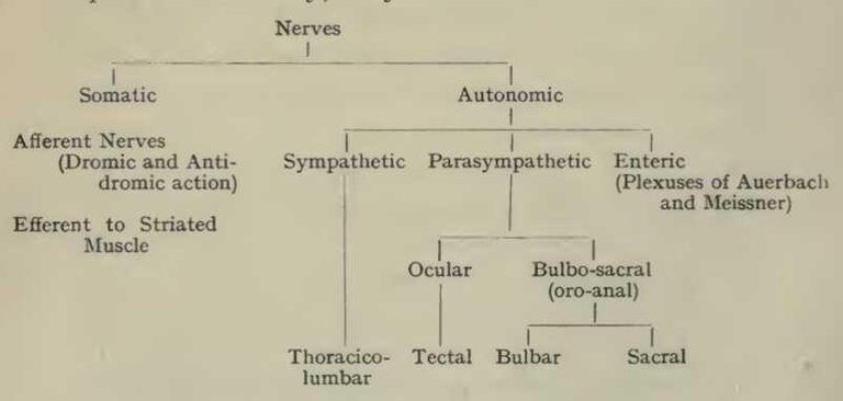
The autonomic nervous system is a part of the peripheral nervous system that controls cardiac muscles, smooth muscles and glands.

It functions largely below the level of consciousness and serves to control visceral activities. The primary target of autonomic nervous system include the following
1.) Circulatory system (heart and blood vessels)
2.) Digestive system (gastrointestinal gland, sphincter, liver and salivary gland)
3.) Reproductive system (uterus and genitals)
4.) Renal system (kidney and sphincters)
5.)visual system (pupil dilator and cilliary muscles)
Comparison of smooth and autonomic nervous system (Differences)
FEATURE
SOMATIC NERVOUS SYSTEM
AUTONOMIC NERVOUS SYSTEM
effectors:
Skeletal muscle.
Smooth,cardiac muscles and glands.
Control:
Voluntary.
Involuntary.
Efferent pathway:
One nerve fibers from central nervous system to effector organ.
Two nerve fibers from central nervous system to effector organ.
Neurotransmitter:
Acetylcholine is its neurotransmitter.
Acetylcholine and non adrenaline is its neurotransmitter.
Effect on target organ:
Has an excitatory effect.
Its effect Could be excitatory or inhibitory.
Effect on denervation:
Placid paralysis
Denervation hypersensitivity
Similarities:
1.) Both the somatic and autonomic nerve are motor and efferent
2.) Both are characterized by afferent and efferent fibers
Divisions Of The Autonomic Nervous System
1.) Sympathetic division:- the sympathetic division functions essentially to adapt the body for physical activity and it does the following
a.) Increases alertness by stimulating the reticular activating system.
b.) Increases blood flow to the cardiac and skeletal muscle.
c.) Reduces blood to the skin and digestive tract.
All this reaction occur during exercise, stretch, danger, competition, trauma, anger, etc.
Physiology And Anatomy Of The Sympathetic Division
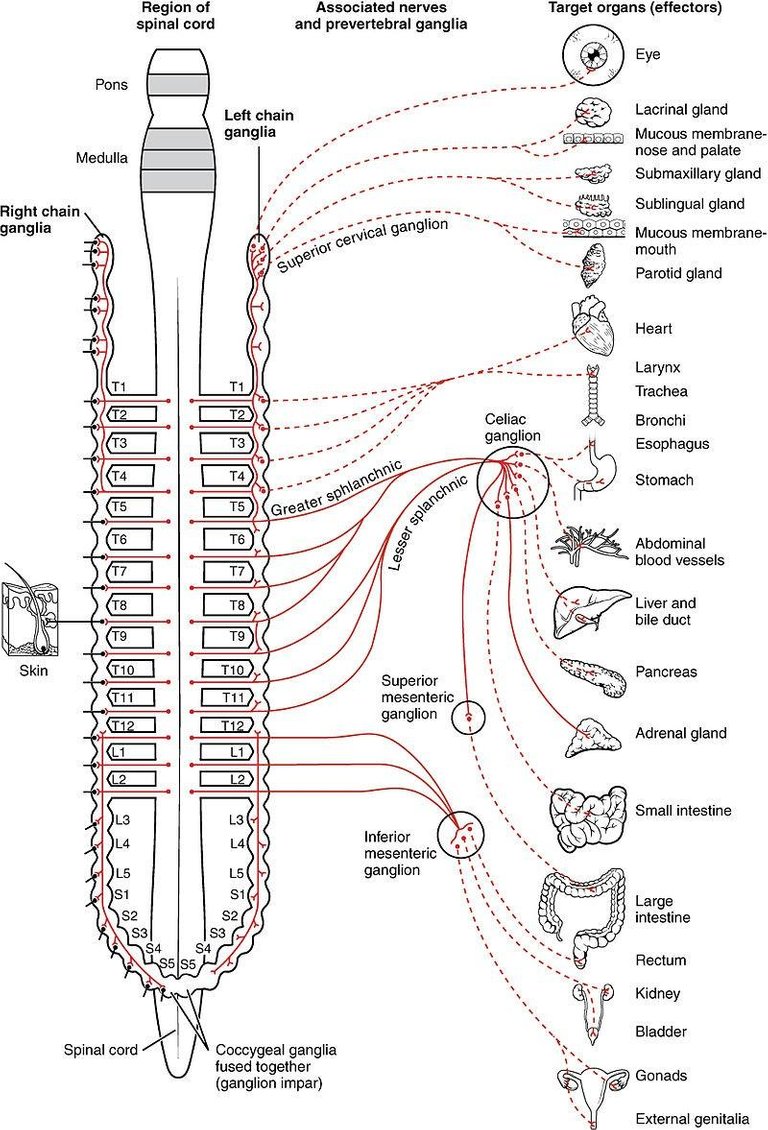
1.) They originate from the thoracic region and lumbar region of the spinal cord. That is they are thoracolumber in origin from t1 to l2.
2.) Their ganglia are described as been para vertebral and collateral. Para vertbral means they are located near the vertebral column. While they are called collateral because they are situated in the thorax, abdomen and pelvis in relation to aorta and its branches.
3.) They have short preganglion fibers and long post ganglionic fiber.
4.) The sympathetic division acts during period of stress or excitement.
5.) Sympathetic effect are catabolic, that is they increase the breakdown process from the body.
6.) They are ergotropic, that is they stimulate the general defense of the body.
7.) They are dynamogenic, that is they are energy producing.
Parasympathetic Division
The parasympathetic division carry out mostly vegetative and restorative function.
Physiologic Anatomy Of Parasympathetic Division
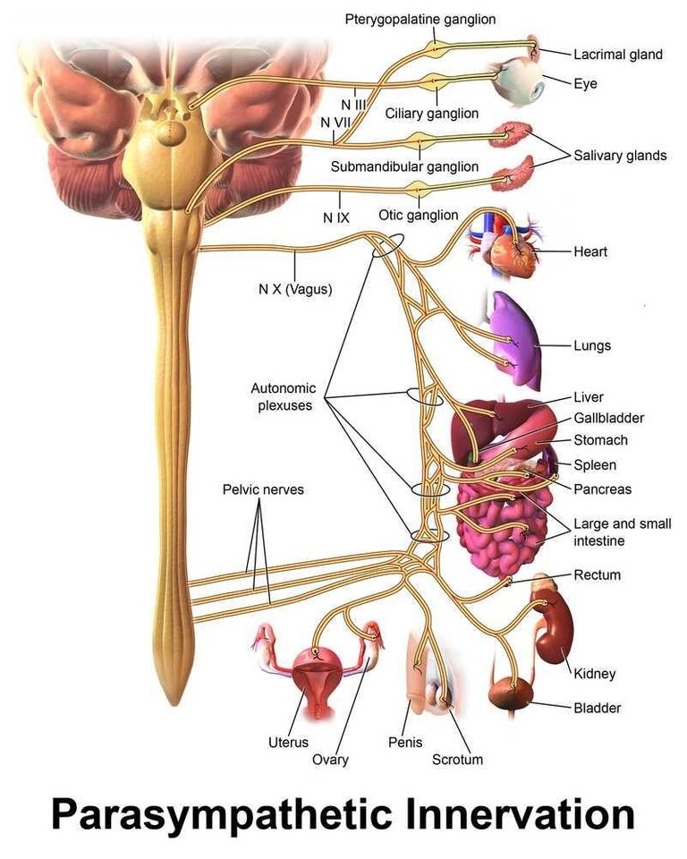
1.) They originate from cranial nerves as well sacral nerves on the sacral area of the spinal cord, that is they are craniosacral in origin.
2.) They have collateral and terminal ganglia:- terminal ganglia are located very close to the effector organ or in the effector organ itself. They have long preganglionic fiber and short post ganglionic fiber.
3.) Functionally, the parasympathetic division acts during quiet condition when the individual is at rest. It makes a homeostatic adjustment necessary for vital function. Note that heart beat, functions of the digestive system and liver, are kept going by the increased activity of parasympathetic division of the autonomic nervous system.
5.) Parasympathetic effect are anabolic
6.) They stimulate the nutritional activities of the body.
7.) They are hypnogenic meaning they produce sleeping or resting conditions.
FEATURE
SYMPATHETIC
PARASYMPATHETIC
origin:
Thoracolumber in origin
Craniosacral in origin
Location of ganglia:
Paravertebral and collateral
Collateral and terminal
Fiber length:
Short preganlionic and long postganglionic fibers
Long preganglionic and short postganglionic fibers
Neural divergence:
It is extensive
It is minimal
Effect on system:
Often wide spread and general
Most specific and local
SIMILARITIES :-
1.) Both enervate mostly same structure
2.) Both cause opposite effect
3.) Both involve multiple receptor system
4.) Both are associated with ganglia
5.) Both are cholinergic at preganglionic synapse
6.) Both are active simultaneously
7.) Both exhibit a background rate of activities called autonomic tone.
ORGANIZATION OF SYMPATHETIC DIVISION
The sympathetic nerves originate from the thoracic and lumber region of the spinal cord, that is from T1 to T2. T1 supplies the head and T2 supplies the neck. T3 - T6 supplies the thorax and upperlimb T7 - T11 supplies the abdomen, T12 - L2 supplies the lower limb. This distribution is only approximate and overlaps greatly. The distribution of sympathetic nerves to each organ is determined partly by the position in the embryo where the organ originates.

ORGANIZATION OF THE PARASYMPATHETIC DIVISION
The parasympathetic nerve is craniosacral in outflow. It consist of cell bodies from the brain stem and sacral part if the spinal cord. From the brain stem we have
Brain stem --- cranial nerve 3
Facial nerve ---- cranial nerve 7
Glossopharyngeal ---- cranial nerve 9
Vagus nerve --- cranial nerve 10
From the sacral spinal cord we have S2 - S4. The vagus nerve is the largest and longest parasympathetic nerve in the body.

AUTONOMIC EFFECT ON VARIOUS TISSUES AND ORGAN OF THE BODY
All smooth muscles and organs of the body receive both sympathetic and parasympathetic innervation. All the life sustaining activities of the body are controlled by sympathetic and parasympathetic division. The balance between sympathetic and parasympathetic division shifts in accordance with the body's changing needs. Neither divisions have universally excitatory or calming effect. For example the sympathetic excites the heart and inhibit digestive and urinary function, while parasympathetic does the opposite. The difference in the neurotransmitters and receptors account for this different effect. The main neurotransmitters of the ANS are :-
1.) Acetylcholine
2.) Non - epimembrane or non - adrenaline.
For all the effect produced by the neurotransmitters released at nerve endings, there are receptors and this receptors include:-
1.) Cholinergic receptors
2.) Adrenergic receptors
Note: nerves that secrete acetylcholine as neurotransmitter are referred to as cholinergic nerve. While nerves that secretes non - adrenaline are referred to as adrenergic nerve. Receptors are responsible for receiving stimulus and making it possible for the nerve transmission. Muscarinic receptors have different sub types which include m1, m2, m3, and m4. And they are found in all effector cells that are stimulated by the postganglionic cholinergic nerves of either sympathetic or parasympathetic nervous system. Nicotinic receptors have 2 subtypes. They are Nm and Nn. They are found in all synapses in the autonomic ganglia. They are found in all synapse in the autonomic ganglia. They are found on cells of the adrenal medulla, again they are found in neuromuscular junction of skeletal muscles as well as points where the preganglionic neuron stimulate post ganglionic cells. Adrenergic receptors are of 2 types alpha and beta. Alpha is subdivided into alpha1 and alpha 2 while beta is subdivided into beta 1, beta 2, and beta 3.
ORGAN THAT CONTRIBUTES TO AUTONOMIC FUNCTION ( ADRENAL MEDULLA )
The adrenal medulla is a part of the adrenal gland situated on the superior pole of the kidney. It is essentially a sympathetic ganglion and consist of modified post ganglionic nerves without dendrites and axon. The adrenal gland and the sympathetic nervous system work together in development and funtion and together are collectively referred to as sympathoadrenal system. When stimulated. The adrenal medulla secretes nor - epinephrine which is about 20% and epinephrine which is about 80% in the blood stream. The secreted nor - epinephrine and epinephrine have the same effect as sympathetic stimulation but they last 5 to 10 times longer. Pulmonary discharge and normal post ganglionic stimulation of sympathetic nerve are collectively known as mass sympathetic discharge.
Reference:
1.)parasympathetic division
2.)neurotransmitters of the autonomic nervous system
3.)wikipedia