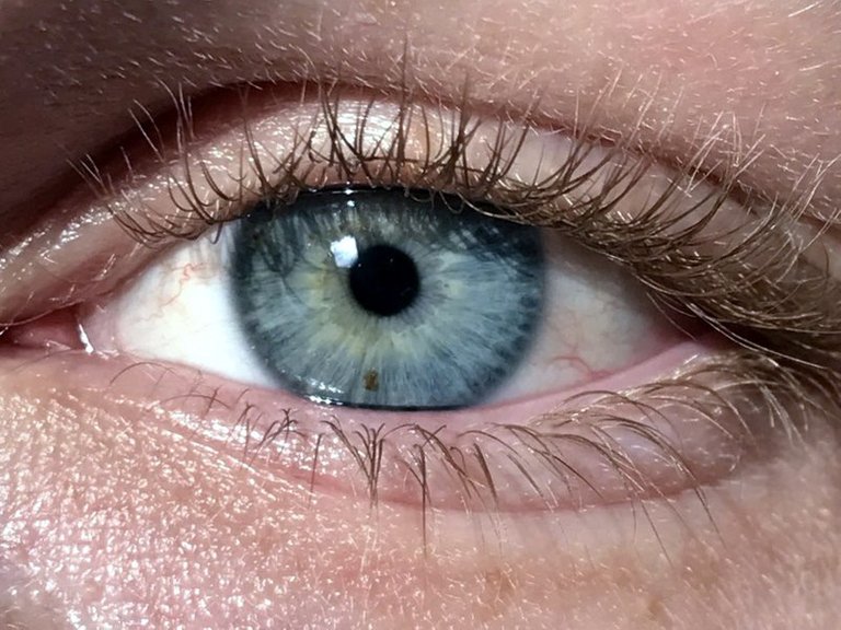
Image used under creative common 4.0
Of all the five senses in human (touch, smell, sight, feel, taste), majority considers the eyes as most valuable and has developed phobia for losing it as they believed the eye is the light of the body irrespective of it size. Hence, Critical examination of its anatomy, mode of operation and possibly health defect is also necessary. The eyes can be regarded as the remote sensor which enables us to see or captures things around us (external environment). Though the eye is a small organ, it’s essentially among the most complex organ of the human body.
THE EYE STRUCTURE.
Its directly located in a bony socket in the skull extending only a small portion outside which can be seen. Scientifically, the wall of the eye comprises of three layers namely: the outermost layer, the middle layer, and the innermost layer.
The outermost layer: It’s also known as the sclera which is a whitish tough layer with tissues connected together. The function of the sclera is mainly to protect and maintains the shape of the eyeball.
The middle layer: also called the choroid is made up of black pigmented cells with very rich supply of blood capillaries, this layers forms the ciliary body and the iris.
The innermost layers: this is the retina; it’s a light sensitive portion of the eye due to the fact that it has numerous photoreceptors which are of two types (rods and cones). The rods are responsible for black and white visions. It’s also helpful to see at night while the cones are responsible for various colour visions. The retina is directly located behind the eyeball. The capillary in the middle choroid supply the retina with available nourishments.
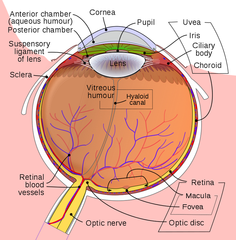
PARTS OF THE EYE

Image used common creative license 3.0
Conjunctiva: the conjunctiva gland is the portion that contains mucus for moistening the eye; it helps to keep the eyes moist at all times. Failure or malfunctions of this gland might probably result to serious pains and itching. The conjunctiva protects the cornea too
Iris: in the iris, the colour of the eye is determined. It’s the segment that gives the eye it colour, it’s the iris that surrounds the pupils in all it sides. The iris widen and shrink the pupils depending on the intensity of light into the eye, if light is low; the iris will widen the pupil and vice versa.
Lens: this is a very transparent layer, after the pupils receives the light from it surrounding, the lens then focus the light unto the retina.
Pupil: it’s at the centre of the eye; it’s like a black dot with tiny hole that allows the passage of light.
Cornea: it’s a domed shaped structure that serves as a cover to the eye against anything which can cause harm to the eye.
Sclera: the sclera is regarded as the outermost part of the eye; it’s white in colour and maintains the shape of the eyeball.
Choroid: it’s the interphone between the retina and sclera which is responsible for the provision of nutrient to other part of the eye.
Retina: it can be found at the back of the eye, it major function is to receive light from the focus and transmit it to electrical impulse before being sent to the brain.
Macula: it’s standing very close to the retina, which helps the eyes to focus on an object.
Optic nerves: this is the collections of nerves that carry impulses from the retina to the brain.
Anterior and posterior chambers: in the eye interior section, the front part is the anterior chamber while the back part is the posterior chamber.
Then how do we see?
Light rays enters our eye when we look at an object, these rays are refracted before getting to the retina. An inverted image is formed the retina which is real and smaller than the object. As the job of the retina is to transmit electrical impulses from the retina to be brain, hence it send impulses to be brain which interprets this image giving it real size and details of object. The above process is very fast and repeated for all objects seen.
Common Eye defects
These are abnormalities in normal functioning of the eye. Examples are myopia, hyper-metropia, astigmatism, cataract, night blindness, conjunctivitis, and xerophthalmia.Below are brief discussion about them.
Xerophthalmia: this eye defects is caused when vitamin A isn’t sufficient in the eye. It’s very possible it leads to blindness.
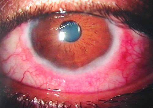
Conjunctivitis: it happens when the conjunctiva is inflamed.

Image used under creative commons license 1.0
Night-blindness: it causes victim not to see clearly at night or when the light is dim.
Cataracts: common to aged people, a condition when the lens becomes cloudy due to advancing age. Using a suitable lens or changing the lens with an artificial one will correct this defect.
Astigmatism: opticians confirmed the presence of this defect in all eye, in some cases the cornea isn’t even thus a distorted/stretched image is formed while some cases horizontal objects are blurred.
Long-sightedness: distanced object are seen clearly while nearby objects appears blurred. Long-sightedness can be best corrected using a convex lens (converging lens).it will converge the image formed behind the retina to be focused on the retina. It’s also known as hyper-metropia.
Short-sightedness: also referred to as myopia. People with this eye defect see near object clearly but not distanced object. A diverging or concave lens is suitable for correcting it.
Care of the eye.
The eye should be cared for very meticulously as it has been considered as the light of the body, we should avoid direct contact with dirty fingers also we should see the optician quickly as soon as we notice any abnormalities in normal functioning of the eye.
References.
Robertsonopt: Parts of the Eye
Wikipedia: Human Eye
Wow! Keep it up. Such a nice post.