G'day team,
This article will require a working understanding of anatomical terminology. If you haven't read my posts on understanding anatomical terminology you can find them here
Anatomy 101 - Anatomical Terminology (Basics)
Anatomy 101 - Anatomical Terminology (Detailed)
Today I thought we'd cover one of my favorite organs, the eyes!
The human eye is among the most heavily evolved and specialized organs on the planet. Many birds may see further, many fish may see more colours and almost all foraging herbivores have a better field of vision. But the human eyes is a master when it comes to distinguishing colours, especially in the range of green, and our visual acuity does well when compared to most animals too.
So... let's get into it.
The Orbit
The orbit is the bony cavity of the skull that holds the eye, it's muscles and other associated structures. The orbit actually gives a great insight into just how complex the structure of the skull is, being somewhat of a junction for many of the bones that make up our facial skeleton and cranial vault.
We won't go into naming all the bones of the orbit in this basic summary, but it is important to understand a few features of the orbit.
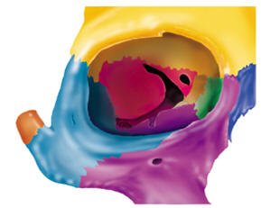
Image source
Optic canal
This is a single hole you can see in the back of the orbit, medially and superior to the other holes. It's through this hole that the optic nerve and retinal artery access the eye.
Superior orbital fissure
This is the superior of the two long holes through the back of the orbit, the shape of which gives it its name 'fissure'. This is an important hole as it's through here that many of the nerves which conduct to the muscles of the eye and face travel.
Inferior orbital fissure
This is actually a continuation of the long hole that forms the superior orbital fissure, but appears at a different angle and transmits far fewer important vessels.
Visual Axis
It's also important to note that from above the axis of the orbits (the directions they point) is not actually straightforward, but outwards at an angle. This means that while we sit here facing forward our eyes are actually both rotated inwards, so as to be focusing on the same object.
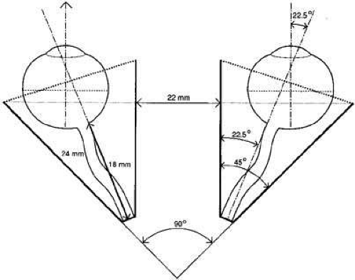
Image source
Now we understand the cavity that they eyes sits in, it's time to have a look at the muscles that surround, and move, the eye.
Muscles of the Eye
Yayyyy muscles! Everyone loves learning about muscles.
Let me change your mind : )
There are six extrinsic (outside) muscles of the eye, which are responsible for moving the eye. These include four straight muscles, and two which attach to the eye at an angle. Let's start with the strait ones:
Superior rectus
Inferior rectus
Lateral rectus
Medial rectus
These muscles all attach to a single fibrous ring sitting at the back of the orbit, and reach forward attaching directly onto the eye.
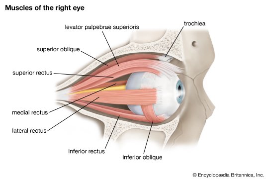
Image source
Each of these muscles is fairly simple. Each pulls the eye in a single direction. But there are two other muscles of the eyes, which are responsible for complex movements which we'll skip over here, but address in the detailed articles.
Superior oblique
The superior oblique muscle starts with the rest of the rectus muscles at the back of the orbit, it reaches forward running superior and medial to the eye (on the inside), it passes over the eye before attaching to a ligamentous loop called the trochlear, before turning around and passing back again to attach to the (here we go!) lateral, superior and posterior aspect of the eye. Complicated? Perhaps a diagram will explain it better!
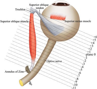
Image source - This is a RIGHT eye, as seen from above
Inferior oblique
Thankfully the inferior oblique is nowhere near as complex! It's the one extrinsic muscle of the eye that starts in front of the eye, medially, and reaches backwards laterally, attaching to the inferior, lateral and posterior aspect of the eye.
Now we understand the muscles that surround and move the eye, we should have a look at the vessels that enter the eye
Vessels of the Eye
There are many important vessels (arteries, veins and nerves) that enter the orbit and supply either the eye or the face, but for the sake of this article we'll just cover three most important ones.
Ophthalmic Artery
The ophthalmic artery branches off one of the main vessels that supplies the brain and enters the eye through the afformentioned optic canal. Right after entering the orbit it branches into a number of smaller arteries, the most important of which is the retinal artery. The retinal artery enters the sheath that surrounds the retinal nerve and goes forward in to the eye where is supplies (you guessed it) the retina.
Optic Nerve
Starting at the eye the optic nerve passes backwards through the optic canal and continues backwards sitting right up under the brain before reaching the optic chiasm, after which it becomes (officially) a part of the brain!
And finally, now we understand everything that surrounds it, let's look at the eye itself!
The Eye
Despite the complexity of it's function the anatomy of the eye is based on a very simple model. In essence the eye has just three layers.
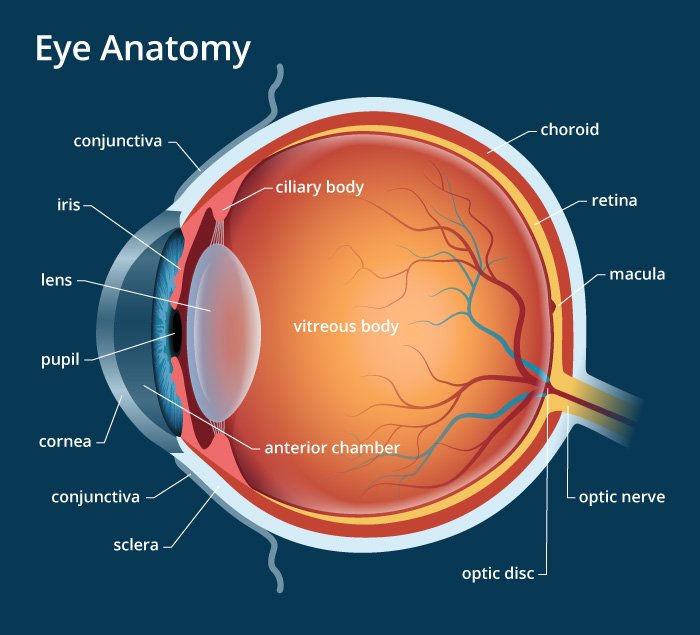
Image source
Sclera and Cornea
This is the outermost, protective, layer of the eye . It surrounds the entire eye and provides protection from insult. Over the lens, where light must pass through, it bulges out slightly and becomes known as the cornea.
Choroid,
This is the layer of the eye that carries both the blood supply and the muscles. Anteriorly this includes the ciliary body, the muscles responsible for changing the shape of the lense when we focus, and the muscles of the iris which change how much light enters the eye.
Retina
This is where all the fancy conversion of light into nerve signals occurs, this is the neural layer that covers the back portion of the eye. There are two basic regions on the retina that are important to note. The first is the macula, which is the centre of the retina and the section that forms our central vision and is particularly sensitive to colour images. The second is the site where the optic nerve leaves the eye, a small section which is not covered in retina and as such a small section at which we are blind. This 'blind spot' can be easily identified with a simple test.
That's All
So that's the eye! Or the basics at least. I'll be posting a detailed article over the next few days which will contain some more (super fun) information on...
- Nerve innervations of the muscles of the eye
- Details of the retina
- Anatomy of the eyelids
- Details of the optic nerve and it's path to carrying information to the brain
Thanks team
-tfc
My Recent Posts
Hi, I'm Tom! (My life of medicine, science and fantasy)
Super Bugs - What is antibiotic resistance and how we are fighting it
Hydatidiform Mole – When Pregnancy Goes Wrong
Tags - Top 20 Profitable, Engaging and Popular tags
New Years Resolutions - How to set goals, and achieve them
Eyes are very interesting. I have some experience in the ophthalmology industry and you get to see how eyes work and what problems they develop.
You're right. They're often one of the hardest organs to work on diagnostically, but their underlying anatomy and physiology is fascinating!
I came to gush about how neat the way that our brain utilizes our eyes to perceive our world, however it appears that I am a post early!
Speaking directly to the anatomy, we dissected cow eyeballs and I was most shocked by how hard and thick the lens of the eye is; I always thought of eyes as squishy. Man I don't miss learning muscles and their insertions though haha
Haha you're right! Nothing worse than rote learning this stuff for the first time. But once you've got it down it feels great!
One more important thing is binocular vision we see two but percieve one image
Yeah you're right. This has so many things that tie into it including the concept of a dominant eye, the reflexive action of yolk muscles and the complex job of the visual cortex to calculate distance based on optical convergence and focus.