Hi everyone, it is always nice to write because I know I have wonderful people who support me through their nice comments and great advice on my previous blog posts. I can't thank you all enough, you spur me. I hope our cordial relationship continues for as long as possible.


Today I was checking out my family’s photo album, it was fun seeing the amazing transformations I've gone through over the years, although my height has been fairly constant since a long time ago. If I asked you to guess the age I first appeared in my family's photo album, I'm sure your answers will range from seven days to six months.
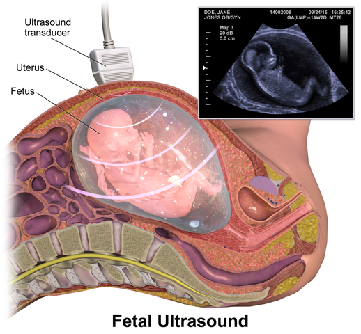
But, I will gladly tell you my first picture isn't close to anything you had in mind, the right answer is seven months. Not seven months old but the seventh month of my mom carrying me in her womb. My first picture is that of when I was still in my mother's womb. I guess you will all now know what I want to talk about. Just so we are on the same wavelength, today I want to talk about using ultrasound in medical imaging.
Like X-ray radiography and magnetic resonance imaging, ultrasound imaging finds wide application in medical imaging. Its most popular use is for checking the condition of a baby in the womb and also in determining the sex of the baby. In normal times, shopping for an unborn child is usually stressful as you don't know the gender of the baby.
You have to get limited stuff and wait till the baby is born which is not so cool as the baby might need some things you have not gotten. Thanks to the technological advancements, ultrasound imaging helps eliminate such problems. Ultrasound scans are not limited to women and unborn babies. Ultrasound is also used to investigate kidney stones or gallstones for men and women.

Ultrasound
The human ear has an upper and lower limit of hearing when a sound wave has a frequency that surpasses the upper hearing limit, such a wave is an ultrasound. It has been concluded that any frequency above 20 kHz is an ultrasound.
As we were taught in physics, longitudinal waves cannot pass through a vacuum, they can only pass through a material. All sound waves are longitudinal waves.
Hence, the speed of an ultrasound depends on the material through which it passes. You may wonder how exactly ultrasounds are produced, I will enlighten you shortly.

How are ultrasounds produced
Waves of high frequency have a short wavelength. These waves are the type used in detecting the smaller features in the body. When an ultrasound scan is required, ultrasonic waves are used. But, how do we get these waves, right?
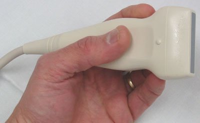
Basically, ultrasonic waves are produced by a transducer. A transducer is an electrical device that changes the form of energy. By changing energy from one form to another, voltage changes and this varying voltage is used to produce ultrasound.
Crystal metals such as quartz are the main component of a transducer. These crystals are known as piezoelectric crystals because when you subject them to a force, they change their shape and produce an electric field. Piezoelectric crystals have an arrangement such that positive and negative ions are aligned side by side.
Ultrasound generally is produced by a vibrating source. Voltage is supplied to the piezoelectric transducer, this applied voltage acts as the vibrating source of the ultrasound waves which is sent through the person taking the scan and the transducer receive the reflected ultrasound waves.
You may wonder as to how possible is it that the transducer sends and also receives signal.
Like a game of squash (when you play alone) you send the ball to the wall, the wall sends it right back at you. Although the returning angle may vary from the one you sent it with, you are the sender and the receiver. This exactly how the transducer works. But, how is it possible, right? The piezoelectric effect works in reverse. That is the simple reason.

Principle of an ultrasound scan
An ultrasound scan is done by simply directing ultrasound waves into the body. If you remember I said earlier that the material through which ultrasounds passes through determines the speed of the wave. So as the waves pass through different tissues, they are reflected differently. These differently reflected waves are put together to form the result of the scan.

If you have had an ultrasound or seen one being done, you know that a gel is applied on the part to be examined. I guess you now know that the principle of an ultrasound scan is in the based on the refraction of waves.
Because air has a poor match of impedance with tissues it creates a reflection and would interfere between the ultrasound waves being sent to through the body as most of the ultrasound will be reflected before reaching its destination. To solve this problem, the gel must be added to the part of the body where the transducer will be placed.
This gel, however, must be of matching impedance with the skin. But, what is an impedance, right? Well, according to technopedia,
Impedance, in electrical devices, refers to the amount of opposition faced by the direct or alternating current when it passes through a conductor component, circuit or system. Impedance is null when current and voltage are constant and thus its value is never zero or null in the case of alternating current.
source
Also, the gel is used as a lubricant, which reduces the friction between the transducer and the skin. It allows the transducer to move freely without pain to the patient.

There are various types of ultrasound scans that can be used, so many that it seems there is a different name for each part of the body that can be scanned. But today I will only be talking about A-scans and B-scans.

A-scan
Following the popular logic that says “as simple as ABC” A-scan is the simplest of all ultrasound scans. Mostly, A-scan is used in determining the length of the eye which can now, in turn, be coupled with the power of the cornea (cornea is the transparent layer and outermost part of the eye) to estimate the correct lenses a person needs.
The process of using A-scan is pretty easy. You only need a transducer, a time-based oscilloscope, and a pulse generator to control the transducer. When all three are connected you can now take your scan.
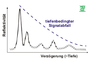
The pulse generator stimulates a surge of ultrasound wave through the transducer and the wave goes through the part of the person you want to scan (where you place the transducer) as the waves hit a tissue, it reflected and detected by the transducer and there is a spike on the screen of the oscilloscope.
The thicker the tissue, the higher the spike is. As the waves travel through tissues they are absorbed and the intensity reduces as the length of travel increases.
If that last statement isn't clear, think of a foam used in cleaning water. If the quantity of water isn't much, only part of the foam will be soaked. So as the quantity of water increases, the more the part of the foam get soaked.

B-scan
If you read my post about X-ray radiography, you will know an advantage CAT scan has over X-rays is its ability to take images from various angles. Well, B-scan is no different. It is just a combination of A-scans from different angles.
To take a B-scan, you need the same equipment used for A-scans with only the addition of sensors. The transducer is moved across the part to be scanned. The wave is reflected and detected by the transducer and is studied to know the length it has travel and what part of the body that represents.
Just like if you are receiving a call from someone in Nigeria, you know the call is from Nigeria because it the number will start with +234. However, knowing the call is from Nigeria might not be enough, you may want to pinpoint the particular place. So when it says they are calling from Ibadan, you know that is Oyo state. So for each part of the body, they reflect ultrasound waves differently.
Instead of spikes appearing on the screen of the oscilloscope, dots are seen when using B-scan. The brightness of the dots depends on the intensity of the reflected ultrasound.

Also, the cost of setting up an ultrasound scan or even taking a scan is considerably less than that of the other imaging technologies. If you take an MRI scan, your result may not be ready for a while depending on the facilities of where you take the scan and who ordered the scan.
But with ultrasounds, you receive the result almost instantly, even some type of ultrasound scan acts like a video recorder as you can see live action of what is going on inside you. Because of this video recorder like feature, ultrasound imaging is used to view how blood flows through vessels.

Conclusion
To improve the quality of the image produced using ultrasound imaging, people who want to take the scan are advised to not drink or eat anything few hours before the scan is done. Although, the advice is for people taking a scan of their digestive system. If you want to take an eye scan, the advice may not apply to you.
There are few issues with ultrasound imaging, the major knock been that ultrasound scan can't be used to detect the most form of cancer. But also, ultrasound imaging is a specialist in a particular area. It is very effective in the detection of very dense breast cancer other form of imaging can't reveal.
The need for an highly skilled operator or a trained professional is another drawback when using ultrasound imaging. Because without them being around, no scan can be done.
Lastly, the main disadvantage of using ultrasound imaging is that it doesn't give an accurate result if the area of interest of the scan is directly under a bone or where the air is. This is why it can't be used to take images of the lungs. The lungs transport air and as explained earlier. The air-tissue reflection will interfere with the ultrasound waves.
Anesthesiology, Angiology (vascular), Cardiology (heart), Emergency medicine, Gastroenterology/Colorectal surgery, Gynecology and obstetrics, Neonatology, Ophthalmology, Pulmonology, Urology (urinary), Musculoskeletal, Scrotal, Nephrology (kidneys) etc are examples of part of the areas where ultrasound scan finds its application. The list of organs that can be scanned is endless. The vast application of ultrasound imaging makes it a very important diagnostic tool in the medical field.
References
Gordon PB. Ultrasound for breast cancer screening and staging. Radiol Clin N Am. 2002;40:431-441
Medical Ultrasound on Wikipedia
Ultrasound
Ultrasound imaging on FDA
If you write STEM (Science, Technology, Engineering, and Mathematics) related posts, consider joining #steemSTEM on steemit chat or discord here. If you are from Nigeria, you may want to include the #stemng tag in your post. You can visit this blog by @stemng for more details.


.png)
Ultrasounds are one vital medical tool. Like you have noted, the fact that it uses no Ionizing radiation as in X-ray imaging is a plus and this makes it safer.
Interesting write up I must say. I like the illustrations you gave, it provided a better understanding of the course.
Best wishes!
Thanks for the kind words
I've came across multiple subjects in the medical field that requires ultrasound to assist in diagnosing many illnesses. The amazing part of this invention is that it is free from radiation unlike most imaging techniques such as CT scan and Xrays. I've seen A and B scans before in the Ophthalmology clinic. Thanks for explaining how the ultrasound works as it helps me a lot in understanding the mechanisms behind it. Great work !
I'm glad I helped you understand.
It's nice to have you in my comments.
Learnt more on Ultrasound imaging usage from this post.. Thanks for making it simple
You are most welcome
You know I thought ultrasound was been mainly used by pregnant women. My bad! I like the topic and the way it's been authored.
Now I can go to the hospital looking at the X-ray machine and the ultrasound's like 'I know who you are and also what you guys do'.
😄
Plot twist, people will be looking at you looking at them like "I don't know you, but I know what you did" 😁😁😁
Lols. Crazy world!
You didn't get the joke
My bad! Maybe. Make it clearer.
Well, great but i don't think my first picture was an ultrasound..
People now do lots of scans now especially the pregnant ones.. But in my time i think it was scare.
Maybe in our country , it finds big application in the investigation of breast cancer.
Interesting article @addempsea. I went for a scan recently, for a slight pain on the right side of my abdomen. I don't know if it was A, B or C scan but it had the video feedback, which the doctor viewed as the transducer moved all over my body. It was an interesting experience. When will they do the colour TV? Things look better in colour.
There are colored ultrasounds already,
These type of ultrasound scans are colored. They are called Doppler ultrasound.
Hi @addempsea!
Your post was upvoted by utopian.io in cooperation with steemstem - supporting knowledge, innovation and technological advancement on the Steem Blockchain.
Contribute to Open Source with utopian.io
Learn how to contribute on our website and join the new open source economy.
Want to chat? Join the Utopian Community on Discord https://discord.gg/h52nFrV
Technology has a way of making life easy. Just like it helps people know the right type of clothes to buy for the unborn child.
Are there cases where the ultrasound can return wrong images? Like, a wrong gender for the unborn baby
Ultrasound provides images, it's up to the radiologist to same what sex it is. And should that be wrong, it's the fault of the radiologist not the ultrasound.
Interesting read. Pls tell me to stop clapping. 👍
This is one of the cases where technology meets medicine.
Despite the stated drawbacks, Ultrasound is of no doubt an essential tool in medical imaging.
Don't stop clapping oh, the more you clap the more my head swells.
Your comment is absolutely spot on!! Thanks for chipping in.
Heard of several scans that were done and changed during birth... Is the ultrasound responsible or the handler?
Great article by the way.
The handler ( a radiologist)
Technology improving medical field through ultrasound. Many round of applause to you on this one sir. It was really simplified and easy to comprehend. Great job mate