Hello everyone, welcome to my blog once again, I am really appreciative of all the lovely comments and nice words I received last time out. Your words spur me on, I hope we can continue our cordial relationship till the end of time.
I suppose y'all by now know that I'm an avid Arsenal FC fan, I've been my whole life, I will be my whole life. In this short time I've spent on earth, arsenal have always had the same manager, a man who go by the name Arsene Wenger, he made his team play beautiful and attractive football. He is everything to the club, in fact, I even thought Arsenal FC was named after him (Arsene Wenger). He's that great of a manager, so when the news reached me that he will be stepping down at the end of the current season, I was gutted, heartbroken and sad. The man that wrote Arsenal FC into folklore will be gone soon. Your invincibles will be forever remembered and your departure will forever leave a hole in my heart. Thank you for all of the memories.
A hole in my heart? Although while this is figurative, it's actually possible to have a hole in your heart. Even if it were true that the hole is in my heart, I'm looking across my chest now there appears not to be a hole there. Who just shouted stupid? Oh the human body is opaque to light so I can't actually see what is happening inside me.
We have learnt in secondary school physics that light does not pass through human body, well other electromagnetic radiation gangsters don't respect us enough not to pass through us. The most popular of the gang is X-rays.
Over the course of this article, I will try and explain to you the physics behind medical imaging using X-ray radiography.

What is medical imaging
Medical imaging is the process and method used when there is need to view human body systems in a way the ordinary eye can not so as to be able to diagnose and track medical conditions. Let's just say medical imaging shows you what you can't see with the naked eyes, like a torn muscle, a cracked bone and so on. There are lots of technologies used in imaging of the human body. The most popular of all I guess is the X-ray radiography, others include thermography, ultrasonography, tomography just to mention a few.
Not all medical imaging technologies actually produce images, some appear in form of electrical signals or graphs, a popular example is the electrocardiography.
I will attempt to walk you through how this technology work. So, grab a chair, sit down, relax and learn.

X-ray Radiography
When I gained admission into the University, the school agreed I was academically fit but to complete my acceptance I had to take a medical examination to determine if I was medically fit for the rigors of what laid ahead. Mere looking at me, you can conclude I'm hale and hearty. I underwent series of tests but one particularly caught my eye, I was to take an image of my chest using X-ray photography. I was confounded by the way the X-ray worked, well not anymore.
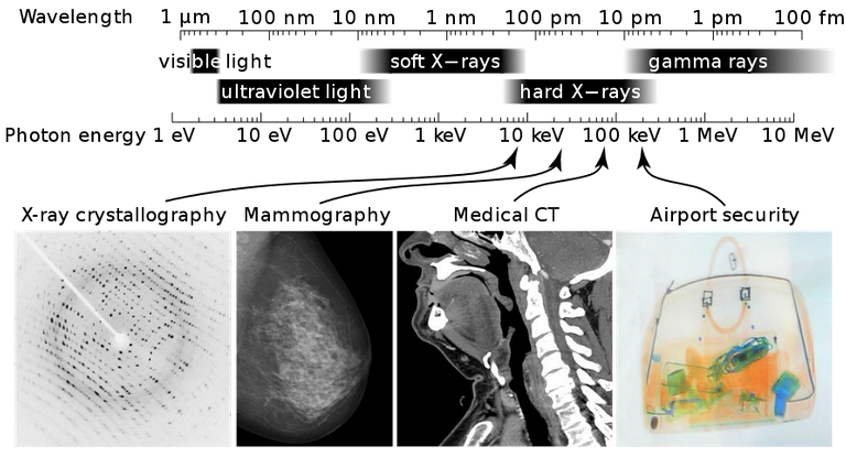
If you check the electromagnetic spectrum, you would find X-rays in between gamma rays and ultraviolet light, X-rays have short wavelengths ( 10-8 m to 10-13 m) and high frequency. They are almost the same with gamma rays, the only difference between them is how they are produced.

Gamma rays are produced when radioactive decay happens, when a nucleus decays, it produces alpha (α) , beta (β) emissions and gamma rays while X-rays are produced when a fast-moving electron is rapidly slowed down. The kinetic energy of this electron as it slows down is transformed into photons of electromagnetic radiation ( X-rays).
X-ray tube
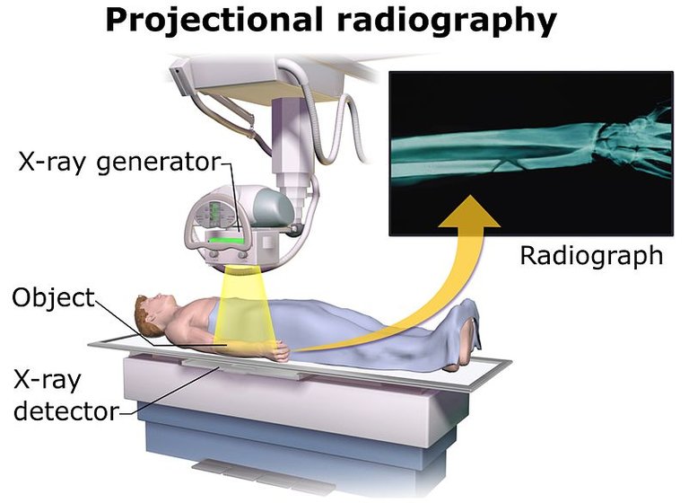
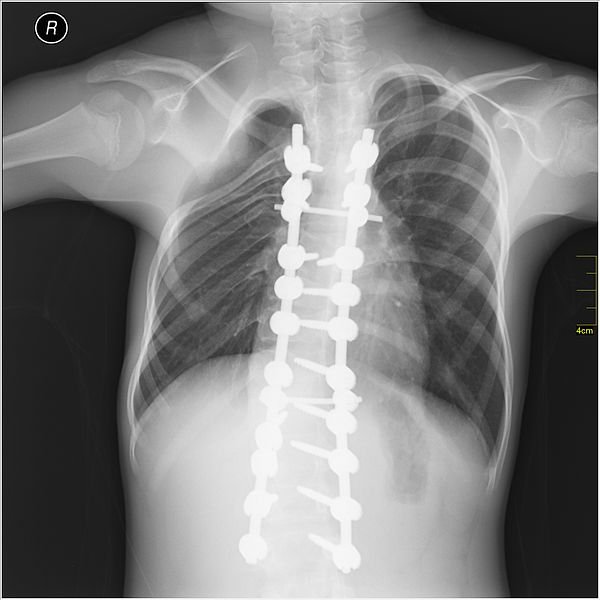
To take the X-ray of my chest, I was asked to stand in between a black sheet placed on the wall and a giant piece of equipment ( I would later find out its called an X-ray tube). Being that I was getting my picture taken I was expecting a flash and I even said cheese. But, I didn't see anything and the woman told me to step out, that we are done here. I almost complained about me not even trying out different poses that I'd learnt in preparation for the photoshoot but the look on her face changed my mind. A few minutes later, she came with my chest in her hand. Not the pretty picture I was expecting, just a white paint brushing on a black sheet. How this had happened I had no idea, I wish I knew then what I know now.

As said earlier, the kinetic energy from rapidly slowed down electrons produce X-rays. A voltage is generated between the anode and cathode by a power source. The voltage injects a stream of electrons across the space between the electrodes. When the accelerated electrons hit the anode, some of its kinetic energy is lost in the form of X-ray particles and scatters in all directions (a basic feature of light waves). X-rays are produced with just about 1% of the kinetic energy of the electrons, the rest is transferred to the anode (which is usually made of hard metal) which explains why the anode is always hot. These X-rays used in medical imaging are called soft X-rays since they have low energy.
Improving on Radiographic imaging
After getting the result of the X-ray of my chest, I took it to school where a doctor examined it and told me it wasn't clear enough, that the person who took the shot clearly doesn't know what she was doing. I went back there series of times, same result until the doctor said I shouldn't go for another month or two. I asked if the result with him was ok, he said no but that I have been exposed to too much of radioactive matter over the past weeks. I had no idea what he was saying but I was happy I didn't have to go to take X-ray pictures for a month.
As a radiographer (someone who takes these shots) you should be concerned with majorly two things, one of which is a radiographer should always strive to improve the sharpness of the images. Lastly, the contrast of the image should be as optimum as possible in order to properly show the tissues and bones.
Improving sharpness
The width of an X-ray beam determines the sharpness with which the image come out. There are factors that determines the width of an X-ray too, the size of the anode for example is one factor as the larger the anode the wider the beam. Another factor that affects sharpness is the size of the aperture at the exit window, the smaller the aperture, the narrower the X-ray beam.
Collimation of X-ray beam is the last factor that affects the sharpness, if some uncollimated beam of X-ray passes through the body and reaches the detector ( the photographic film), they will cause fogginess of the image and reduce it's sharpness. An anti-scatter screen can be used to absorb these uncollimated beam of X-ray, this screen can be made of materials such as lead that are opaque to X-rays, the lead absorb the uncollimated beam of X-ray.
Improving the contrast
To say a contrast is good is to say there is a very clear distinction between the blackening of the photographic film as the X-ray passes through separate bones and tissues. What really determines the contrast of an X-ray is its hardness.
If you break a bone and you want to find out to what extent the bone broke, you will have to take an X-ray. We all know this right, but what you don't know is the type of X-ray that will be used. Since the bone is an excellent absorber of radiation, hard X-rays are used in the imaging process.
But if it were say a person has breast cancer and is to take an X-ray, soft X-rays are used because unlike bones, tissue (the breast is made up of entirely tissues) are very poor absorbers of radiation. Using soft X-rays also equals longer exposure time.
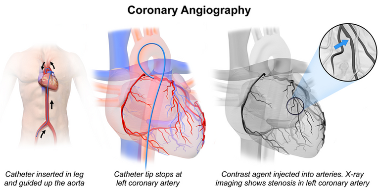
As stated earlier, longer exposure time is hazardous to the health. So how then do we take images of tissues without having to be exposed for so long. This is where contrast media come in play. Contrast media are substances which are good absorbers of X-rays examples include iodine and barium. A person requiring a X-ray scan of his or her tissues will have to ingest a barium or iodine filled meal or have it injected to the area(tissues) to be scanned. The tissues then becomes a good absorber of X-ray for which hard X-rays can now be used.

Risks associated with X-ray
Ionising radiations can destroy living tissues, this destruction leads to mutation which in turn can lead to the growth of cancerous tissues. It is of prime importance to keep the degree of exposure to a minimum.
Although to record X-ray images, it can be done using photographic film or digitally. It has been said that photographic film are weak absorbers of X-rays so you have to be exposed for longer time with increasing intensity. To avoid exposing people to too much of radiation, intensifier screens are used. This is like using the microphone in which you feed in little sound and it magnifies it. Well for intensifier screens, you feed in a X-ray and it magnifies it without increasing the intensity of the radiation.
Image intensifiers are used in digital systems, a visible light is produced when X-rays strike a phosphor screen. Unlike in photographic film where electrons hit the anode to produce X-rays. In digital systems, when electrons are accelerated between the positively charged anode so they can strike a screen, visible light photons are produced. The images on the screen is viewed on a television and can as well be stored electronically. An advantage digital systems have over photographic films is that it can be easily stored and shared.

Limitations of X-ray radiography
The major limitation the X-ray radiography has is that it is a 2-D imaging system. What do I mean by 2-D? When you take an X-ray of a body most of the body parts are superimposed on each other. It won't be easy for one to know what bone is what since they are overlapping. However this problem can be solved using a CAT(not that one) scanner, wherein you take pictures at varying angles and can now see everything that's happening with the body, the bones are not overlapping no more cause you have the knowledge of the bones from different angles now.

Conclusion
Imagine everytime you broke a bone, your skin has to be torn to see the extent of the damage. I can’t even imagine the pain even if painkillers were administered. X-ray radiography helps you in that regard as you won't have to undergo the gruesome process of cutting your skin. X-rays are definitely live savers.
Although they save lives, too much of their radiation can also cut a life short. They also increase one’s chances of developing cancer. Women are more susceptible to developing cancer even when they receive same amount of radiation with men. I even read some doctors because of financial gains subject patients to repeated radiation exposure.
In an attempt to reduce the amount of radiation we are exposed to, doctors have been urged to only recommend X-ray examinations only when the are absolutely sure it is needed and when it is sure it will help in the diagnosing and treating of a medical condition.
Should you have to take an X-ray, there are standard questions the doctor should ask you before issuing an X-ray examination for you. The first question should be about the last time you had one, another (if you are a woman) should be if you are pregnant. Also in an attempt to make sure your doctor knows what he/she is doing you too can ask him questions like how the result will be used in evaluating your present predicament.
References
Krestel, Erich (1990). Imaging Systems for Medical Diagnostics. Berlin and Munich: Siemens Aktiengesellschaft. pp. 318–327.
If you write STEM (Science, Technology, Engineering, and Mathematics) related posts, consider joining #steemSTEM on steemit chat or discord here. If you are from Nigeria, you may want to include the #stemng tag in your post. You can visit this blog by @stemng for more details.


Enlightening!!
Xrays find a vast application in the medical field, however, limited to, though the risks are scary but not one we should fret about right?
Overall Great work done boss!!!
Should you not be exposed for too long, you have nothing to fret over.
I Will like to peep in on what you asked. If one is expose to too much radiation though i don't know how often when calculated can cause aplastic aneamia (this is when the body can no longer produce new red blood cells. Though it mostly rare) as radium is not a friend of the body.. Very crazy element i must say...
and i also heard that xray should be done maximum of 5 times in a life time. Although i don't know how true this is.
The five time thing is a lie, I guess it's all about the amount of radiation.
Hello! I find your post valuable for the wafrica community! Thanks for the great post! @wafrica is now following you! ALWAYs follow @wafrica and use the wafrica tag!
Thanks for educating us about X-ray. I've had it just once and I pray I don't have cause of having X-ray done often as it comes with some risk..Beautiful technology though! Thanks for sharing.
I hope you don't have it again too.
Thanks for stopping by.
I have only done an X-ray once in my life and it wasnt as painful as i thought it would be (it wasn't at all)
nicely written article
I've never had it though, other than that time the school asked us, guess I'm one of the lucky ones.
Your comment is much appreciated.
This article is pretty educative as well as informational. I like the part where you explain the cancer-causing capability of x-ray. That is why I hate going for an x-ray, I do not want anything ionising my precious tissues :)
LoL.... You only have to take X-ray when it is absolutely necessary. Other than that your precious tissues are very safe.
Absolute simplicity!
I love how you took us through the whole technology, it's usefulness and limitation. You sure did your research well..
Well, till now, I've only had one and I was able to know that it's not painful as I always thought..But definitely, I don't want to have another...It's wierd!
Thanks for this awesome post..
It delights me to know that you enjoyed the post.
Your nice words are comforting and thank you for stopping by.
Always a pleasure...
Hard and soft X-rays? Never knew there are types of X-rays, besides I never thought all that much is done other than taking the image, no wonder some hospitals take hours before it is ready, in fact some will require you to return the next day.
While I was reading the part where you discussed d danger of excess exposure, I remembered seeing an episode of "A thousand ways to die" where a doctor for a crazy reason left his patient exposed for too long, and the patient also didn't leave the bed for the same crazy reason until the rays fried him.
I've both enjoyed and benefitted greatly from this article. Bravo
X-ray imaging should be like flash running past the sun, and when you decide to walk like a turtle in front of the sun, I guess you know what will happen.
Thanks for the compliments.
I once took an x-ray i thought it was going to be painful i almost fell off the block when I heard the switch come on.. Then suddenly i was asked to get up... I was like was it done??
Only for the woman to look at me smiling. I also heard it could cause cancer. So i am not really a fan of it.
I cant imagine them having to tear me apart to take a look.
Knowledge is key.
Thanks for dropping by.
Kudos to you on this. Sincerely i never knew it had types even with my little knowledge of x-rays.. Hard and soft indeed(laughs).
I don't have anything else to say than i am glad i read this post. Very informative !
Thanks brother. I very much appreciate your comment.
I love the way you have crafted the article, explaining the whole thing with your personal experience.
Thumbs up man!
I'm glad you enjoyed it.
It's always nice to see you in my comments.
However, X-rays will never allow for the observation of the hole in your heart as heart is not a bone :D
A contrast media can be injected into my heart. 😂 😂 😂
It's nice having you in my comments
Please don't do that :D
I won't have a hole in my heart.
Good! :)
Congratulationsdaily compilation 256Gems! We strive for transparency., your post was discovered and featured by @OCD in its ! You can follow @ocd – learn more about the project and see other
here.If you would like your posts to be resteemed by @ocd to reach a bigger audience, use the tag #ocd-resteem. You can read about it
SteemConnect or on Steemit Witnesses to help support other undervalued authors!@ocd now has a witness. You can vote for @ocd-witness with
I must say what you do not know would stay unknown. I have had X-ray twice now, one for school and the other for NYSC (not compulsory tho), and the experience in the physical was awesome.
The aftermath of getting too exposed to the rays as highlighted in your post is frightening. Therefore, I must guard the result of the previous one well. 😆
Thank you once again.
Thank you too.
Sweet! i've really learnt a lot about x-ray, although i can't relate cos i've never had one.
Thanks for putting this out, and i must confess, i enjoyed your style of presenting it. I thought i'd get bored reading cos of its length, but the reverse was the case☺
I'm glad I didn't bore you with my long post.
Thanks for the lovely comment.
My own view about exposure to x-tray is just that too much of it is not good and could result to short life and should only be recommended when it is necessary.
My own view about exposure to x-tray is just that too much of it is not good and could result to short life and should only be recommended when it is necessary.
Thanks for the additional information.
Congratulations @addempsea! You have completed some achievement on Steemit and have been rewarded with new badge(s) :
Click on any badge to view your own Board of Honor on SteemitBoard.
For more information about SteemitBoard, click here
If you no longer want to receive notifications, reply to this comment with the word
STOPHow do they collimate the beams?
You can collimate beam by using metal tubes to absorb X-rays. Which in turn produces a parallel-sided beam which is collimated.